|
Deutsch English Home Me My Work My Hobbies My Links Perl-Download Zone ITEM-Algorithm My Papers |
WorkMy work at the biophysical group within the center for medical physics and technology at the university of Erlangen-Nürnberg deals with the investigations on the force generation of cells. Cell-mechanics deals with the investigation on force generation of cells during movement and the stability of cells. It is a field of research between physics and biology. Cells at rest provide stability against external forces by means of their cell skeleton. This consists of mikrotubuli and actin-filaments (only mentioning the ones we are interested in). Especially the actin-filaments are highly dynamic construction and can be changed on the timescale of minutes to react on external forces. One not yet solved question is how the cell gets this mechanical feedback (mechanotransduction) and how the cell regulates the changes in the filament-structure. To investigate these question we observe the cells during conditions where the cell develops force: either by means of external drugs that act on the skeleton itself or by letting the cells invade into a gel. This invasion is tracked by a videocamera mounted on top a microscope. Talking stacks of images one gets the possibility to reconstruct the forces generated by the cells by tracking beads embedded within the gel. Formerly I worked at the "Physikalisches Institut": My work at the university of Erlangen-Nürnberg deals with the Compton-Camera. The Compton-Camera is a medical nuclear imaging device. In contrast to the well known X-ray tubes, where the "shadow" of the patient is registered, ITEM is out of the family of functional imaging techniques. There the patient "glooms" himself: By applying (lightly) nuclear active substances (in collection with a biological substance called tracer) into the patient the task is to localise the position of tracer accumulation. Dependent of the tracer substance one can
Even so sounding quite simple it is not. There are several techniques trying to do the trick: PET (positron-emission tomography), SPECT (single photon emission computed tomography) and of course Compton-Camera. PET is somehow little different, so let's skip this one and let me explain how SPECT works: The external detector is shielded from the radiating patient in that sense, that only radiation coming right straight upto the detector can reach it and results in a signal. Then one known at least the line of sight and it is possible to do an image reconstructing by looking at the patient from several different positions. 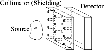 The big disadvantage is: Lots of the radiation leaving the patient does not reach the detector. (This happens almost always when the radiation does not come to the detector in a straight line. Remember ?:-) So one has to apply lots of radiation to the patient to achieve a image. (Lots is not really a lot. This is because the reconstruction methods work pretty well, so one can get good results with poor data. So the dose (measuring how many "decays" the patient had to suffer) is similar to one from a typical CT (Computed Tomography) image, or transatlantic flight, or working in a house made of sandstone, or ... In other words: Lots is not LOT, but it could be less.) This is where the Compton-Camera enters the field: The basic idea is to get rid of the shielding (collimation) by substituting this one by another detector plane. Then the particle can interact in the first detector-plane and gets scattered in the second one. 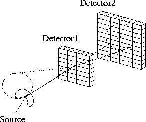 So in principle it is possible to reconstruct where the radiation cane from. (The interaction has to be a Compton-scattering; this is where the camera's name comes from.) In practice this is not so easy and one can only reconstruct the position up to a cone ambiguity. The hope is now that the dose reduction achievable by a Compton-camera is a really big deal (up to 100 times more efficient than a SPECT), but the hardware as well as the reconstruction software (algorithms) are quite difficult. So my work is to improve the Compton-camera by simulating several design and most of all do a image-reconstructing. Being quite difficult this lead to a complete new kind of image reconstruction algorithm. |